Micro-PIV
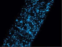
Microscopic PIV Image
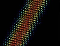
Resulting Vector-Field
The light source is typically a double pulsed Nd:YAG or a cw laser that is coupled through an optical fiber into a fluorescence microscope and focused with a high numerical aperture on a microfluidic device.
The microflow is seeded with fluorescent particles. A microscope lens collects the particle signal that has a longer wavelength than the illuminating light.
This signal is separated from the laser light by a filter cube and is recorded by a double-frame CCD camera like the Imager Intense. The double frame images are evaluated with conventional PIV algorithms.